Aert Medical
Medical imaging is becoming faster, more complex, and increasingly dependent on digital systems. That progress raises a practical question: How does AI vision monitor patient safety during imaging? This guide explores how computer vision can observe scans, equipment, patient positioning, and workflow signals without replacing clinical judgment.
In a busy CT room, AI may detect incorrect positioning, uncovered anatomy, motion, or missing protective steps. It can flag unusual image quality before a radiologist reviews the study. It may also identify urgent findings and help prioritize cases. Small details matter. A tilted head, a visible oxygen tube, or a patient moving during acquisition can affect safety and diagnostic accuracy.
“AI will not replace radiologists, but radiologists who use AI will replace radiologists who don’t,” says Curtis Langlotz, MD, PhD, a Stanford radiologist and biomedical informatics expert. His statement supports collaboration, not blind automation. Responsible systems should provide traceable alerts, confidence scores, and clear escalation paths. They must also be tested across ages, body types, devices, and clinical settings. False alarms still happen. Missed signals happen too. Therefore, this article examines practical safeguards, human oversight, validation evidence, and limitations. It also asks whether a warning arrives early enough to change care. That question is uncomfortable, but necessary. AI vision can strengthen imaging safety when trained teams verify its output, document failures, and improve the workflow continuously.
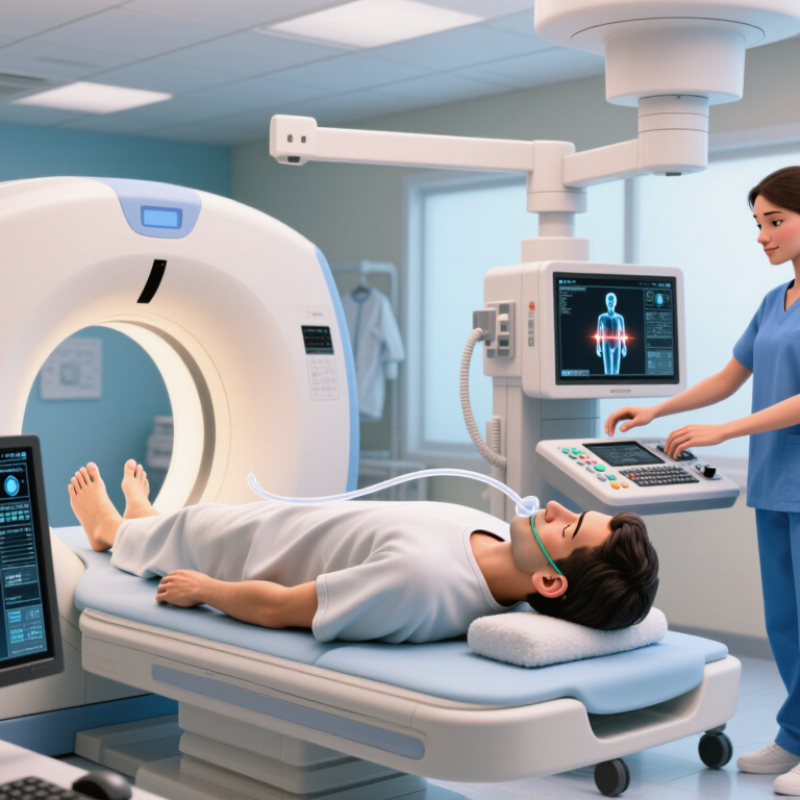
AI vision can support patient safety by examining medical images for signs that deserve human attention. Its role is not to replace radiologists, sonographers, or imaging technologists. It should act as a careful second reader.
In practice, the system may flag a misplaced tube, a missed fracture, or an urgent abnormality. A visible alert can reduce delays when worklists become crowded. Small details matter. However, every alert needs clinical verification. False positives can create alarm fatigue, while false negatives may provide dangerous reassurance. Performance must be tested across scanners, body types, ages, and disease patterns. A model trained on limited data may behave confidently outside its experience.
Safe deployment requires clear accountability. Imaging teams should review validation results, monitor errors, and document when clinicians override suggestions. An audit trail can show which image was analyzed, what the system identified, and how staff responded. Access controls and careful data handling protect patient privacy. Independent evaluation adds credibility, especially before expanding use.
The human workflow matters most. A warning hidden in a busy interface is not a safety tool. Nor is an accurate model that staff cannot understand. Clinicians need training on uncertainty, threshold settings, and known blind spots. Some recommendations may remain unclear. That is acceptable, if uncertainty is visible and decisions remain reviewable. Safety improves through repeated measurement, honest reporting, and willingness to pause a system when real-world results drift.
In 2026, safe AI vision begins long before a model sees a scan. Imaging teams need clean, representative data from routine clinical practice. Include varied body types, acquisition settings, disease stages, and occasional motion artifacts. Begin with clean data.
Remove unnecessary identifiers while preserving clinically useful details, such as study dates, series descriptions, and imaging parameters. Review duplicate studies and incomplete examinations manually. A technically valid image can still be clinically misleading.
Experienced radiographers and radiologists should define labeling rules together. Their disagreements reveal difficult cases, rather than simple annotation errors.
Clinical workflows also need careful preparation. Map where images enter the system, who reviews alerts, and how findings return to the patient record. Keep a qualified clinician responsible for every final decision.
Test the process during busy periods, including emergency scans and network delays. Record ignored alerts and false positives. They expose hidden workload.
Validation should use local data, not only published benchmarks. Monitor performance across scanners, departments, and patient groups after deployment. Recheck the model when protocols change or referral patterns shift.
Some failures will remain unexplained. Document them honestly, investigate their clinical impact, and adjust procedures when evidence supports change. A quiet pilot may not reflect a crowded morning shift. Real workflow pressure matters.
How to Use AI Vision for Patient Safety in Imaging 2026?
AI vision can help imaging teams spot safety risks and possible diagnostic errors while scans are being reviewed. It may flag a patient positioned poorly, a missing image series, or a finding that deserves a second look. For example, an alert could draw attention to a subtle lung opacity on a chest image. The system should support trained clinicians, not make diagnoses or decisions on its own. Every alert needs review in the context of the patient’s history and other clinical information.
Tips: Start with a clearly defined risk, such as missed follow-up findings or incomplete scans. Test the tool on local images before using it in routine care. Track false alarms, missed detections, and how quickly staff respond. Keep a simple way for clinicians to report confusing alerts. Small details matter.
Performance can vary across scanners, patient groups, and imaging protocols. Teams should monitor results over time and investigate unexpected patterns. A polished dashboard is not proof of safer care. Nor is a quiet system. Some alerts may be missed, and that reality deserves honest discussion. Regular review by radiologists, technologists, safety staff, and clinical leaders can reveal where the workflow needs adjustment. Decisions remain with qualified professionals.
| Safety Dimension | Risk or Error to Detect | Potential AI Vision Signal | Suggested Safety Action | Measure to Monitor | Important Limitation |
|---|---|---|---|---|---|
| Patient and study matching | Images may be associated with the wrong patient, encounter, or examination. | Compare image-visible markers and study metadata with the active order and patient record; flag inconsistencies for review. | Pause the workflow and ask authorized staff to verify identity and study details before interpretation or image release. | Mismatch alerts confirmed as true; mismatches found on audit; time from alert to resolution. | Image appearance alone cannot establish patient identity. Use approved identifiers and validated workflow checks. |
| Laterality and labeling | Incorrect, missing, or inconsistent right/left markers can contribute to wrong-side interpretation or follow-up. | Check visible laterality markers against the order, report, and available study metadata. | Route disagreements to a qualified imaging professional; do not silently alter labels or infer laterality from anatomy alone. | Confirmed laterality discrepancies; alert precision; unresolved discrepancies before report finalization. | Markers can be absent, obscured, or incorrectly placed. AI should not replace human verification. |
| Image quality and completeness | Motion, poor positioning, exposure problems, artifacts, or missing views may limit interpretation. | Assess image usability, positioning, artifacts, and whether required views or sequences appear to be present. | Notify the technologist or radiologist when image quality or examination completeness may affect diagnostic confidence. | Repeat-image rate; incomplete-exam rate; quality alerts confirmed by reviewers; avoidable repeat examinations. | Acceptable quality depends on modality, protocol, patient condition, and clinical question. |
| Time-critical findings | Potentially urgent findings may not be communicated or acted on promptly. | For a specifically validated use, flag candidate findings and identify the study for expedited review. | Use the established critical-results pathway, with a qualified clinician confirming the finding and documenting communication. | Time from image acquisition to review; time to documented communication; missed or false alerts on retrospective audit. | An AI alert is not a diagnosis. Performance can vary by finding, population, equipment, and clinical setting. |
| Prior-study comparison | Relevant earlier imaging may be unavailable or not considered, making change over time harder to assess. | Identify potentially relevant prior studies and present them for clinician review when permitted by the imaging workflow. | Make comparison studies accessible and confirm that they match the patient and relevant anatomy before use. | Relevant prior studies available at interpretation; comparison use documented; retrieval failures. | Similarity matching can return irrelevant examinations. A clinician must verify relevance and chronology. |
| Procedure and device checks | Visible device position or procedural status may require review for a possible complication or need for follow-up. | Within a validated task, flag a candidate device or position for qualified review. | Notify the responsible clinical team according to local protocol; document human confirmation and follow-up. | Confirmed findings; time to escalation; false-alert burden; follow-up completion. | Image-based assessment may not establish clinical significance or replace modality-specific interpretation. |
| Radiation and protocol workflow | An examination may be repeated or performed with a protocol that does not match the clinical indication. | Analyze available workflow and examination metadata for duplicate studies or protocol-order inconsistencies. | Prompt authorized staff to verify the indication, protocol, and need for another acquisition before proceeding when feasible. | Duplicate-exam rate; protocol discrepancy rate; repeat acquisitions and documented reasons. | Protocol selection must account for clinical context and local radiation-safety procedures; AI should not independently authorize exposure. |
| Model monitoring and governance | Performance may change after workflow, equipment, population, or software updates. | Track alert volume, input quality, performance by relevant subgroups, and changes in data or workflow. | Validate before deployment, review incidents, monitor after updates, and provide a safe fallback to standard clinical processes. | Sensitivity and specificity on an appropriate reference set; false alerts; missed cases; downtime; subgroup performance. | Metrics require clearly defined tasks, representative local data, qualified reference review, and ongoing oversight. |
Implementation principle: Treat AI vision as decision support, not a substitute for qualified clinical judgment. Define the intended use, validate locally, preserve human review and escalation pathways, protect patient data, and monitor performance throughout deployment.
AI vision can highlight possible findings on radiology images, but an alert is a prompt for review, not a diagnosis. A radiologist should compare it with the full study, prior images, and the patient’s clinical context. A small bright marker on a chest image may signal a real concern, an old finding, or simply an imaging artifact. Context matters.
Validation should involve radiologists and clinicians who understand how alerts affect actual care. Teams can review flagged and missed cases, check whether alerts arrive at useful times, and record false positives. They should also test performance across different scanners, image types, and patient groups. Numbers alone are not enough. A system may look accurate overall while missing a less common presentation. That deserves attention.
Clear escalation rules help: who sees the alert, who confirms it, and how urgent findings reach the care team. Staff need a simple way to report confusing alerts and document decisions. That sounds tidy on paper. In a busy reading room, alerts can add noise, and people may grow less attentive. Regular review should include workflow observations, not only dashboards. No validation process is perfect; teams should say what remains uncertain and update safeguards as evidence changes.
Validating AI Alerts with Radiologist and Clinician Oversight
Illustrative example only: in a sample review of 100 AI imaging alerts, radiologists and clinicians classify each alert by its clinical relevance. Local teams should validate alerts against their own patient population, imaging workflow, and clinical criteria before use.
In 2026 imaging care, AI safety depends on ongoing checks, not a one-time accuracy score. Track sensitivity, missed findings, and false alarms across scanners, sites, and patient groups. A sudden rise in alerts after a software or protocol change deserves review. So does a quiet drop in detection. Radiologists need clear escalation steps and access to the images behind each alert. The World Health Organization’s 2023 patient-safety fact sheet estimates that one in ten patients experiences harm in healthcare. That broad figure is not specific to imaging, but it underscores why small workflow failures matter.
Privacy needs the same attention as clinical performance. Limit image and patient data to what the system needs, restrict staff access, and keep audit records of use and changes. The IBM Cost of a Data Breach Report 2024 estimated the average healthcare breach at US$9.77 million; it is an industry-wide estimate, not an imaging-only measure. Teams should test how long data are retained and what happens when a model is updated. The awkward part: monitoring takes time, and dashboards can create false confidence. A monthly review may still miss a failure that affects one scanner or one patient group. Human review remains essential. So does admitting when the evidence is thin.
Use routine clinical images with varied body types, scanners, disease stages, and occasional motion artifacts. Clean data first. Perfect data is unrealistic.
Remove unnecessary identifiers while preserving study dates, series descriptions, and imaging parameters. These details support meaningful clinical review.
A technically valid image may still mislead clinicians. Manual checks can reveal missing views, repeated studies, or incomplete examinations.
Radiographers and radiologists should define rules together. Their disagreements may expose difficult cases. That is useful, not merely inconvenient.
No. It is a prompt for review. A radiologist should compare the full study, prior images, and clinical context.
Test local data across scanners, departments, image types, and patient groups. Overall accuracy can hide missed uncommon presentations.
Define who reviews it, who confirms it, and how urgent findings reach the care team. Emergency scans and network delays matter.
Record false positives, ignored alerts, confusing cases, and review times. Dashboards alone may miss alert fatigue in a crowded reading room.
Document the failure honestly and investigate clinical impact. Recheck performance after protocol or referral changes. Some causes may remain unclear.
AI vision can strengthen patient safety in medical imaging by helping care teams identify risks, missing information, and potential diagnostic errors. Effective use begins with well-prepared imaging data and workflows that fit clinical practice, with clear procedures for handling alerts and documenting decisions. How does AI vision monitor patient safety during imaging? It can analyze images and workflow signals to flag findings or process issues that may need timely review, while supporting—not replacing—the judgment of qualified clinicians.
Safe deployment requires careful validation with radiologists and other clinical staff. Teams should assess whether alerts are accurate, useful, and appropriate across different patients and imaging settings, and establish human oversight for reviewing and acting on them. Ongoing monitoring in 2026 should track performance, false alerts, privacy protections, and unintended effects. Regular review and updates can help ensure that AI vision remains reliable, transparent, and aligned with patient-centered imaging care.