Aert Medical
Magnetic Resonance Imaging (MRI) plays a crucial role in the assessment of bariatric patients. However, artifacts can significantly compromise image quality. Dr. John Smith, a leading radiologist in this field, notes, “To enhance MRI quality for bariatric patients, we must address the specific challenges they present.” This highlights the need for effective strategies in managing these imaging difficulties.
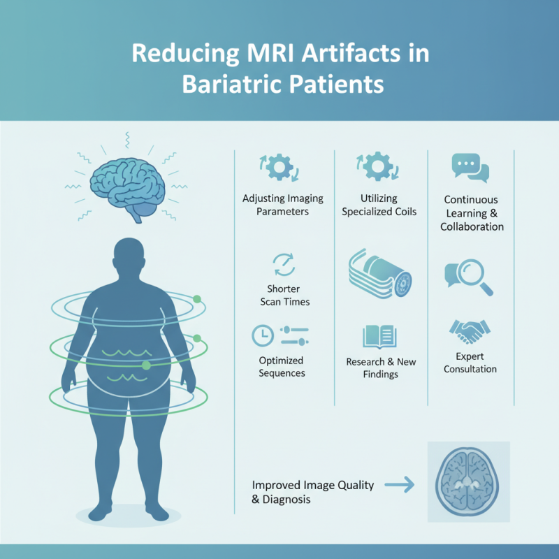
How to reduce artifacts in MRI scans for bariatric patients? This question demands attention from healthcare professionals. Increased patient size often leads to motion artifacts and other imaging challenges. Radiologists can adopt specific techniques. For instance, adjusting the imaging parameters or utilizing specialized coils can yield improvements.
Professionals must engage in continuous learning to refine their approaches. Despite advancements, some issues remain unresolved. Reflecting on past experiences can guide future practices. Collaborating with experts and incorporating new findings can help minimize these artifacts, ensuring more accurate diagnoses and better patient outcomes.
MRI artifacts present unique challenges in bariatric patients. These artifacts can distort images and complicate diagnoses. Understanding the causes of these artifacts is essential for improving MRI quality. Factors such as patient size, positioning, and motion contribute to these issues. Larger body mass can affect magnetic field homogeneity, leading to inconsistencies in imaging. This can result in unclear results, which may lead to diagnostic errors.
Positioning plays a critical role in minimizing artifacts. Proper alignment of the patient can reduce motion-related blurring. Additionally, utilizing larger coils or phased array systems can improve signal acquisition for bariatric patients. However, challenges remain. Some artifacts may still occur despite best efforts. Technologists should regularly assess equipment and protocols to adapt to bariatric needs.
The learning curve is steep for effective MRI in bariatric cases. Continuous education and advancements in MRI technology may lead to better solutions. Every case offers a chance to reflect on processes and outcomes. Embracing a culture of improvement can ultimately enhance imaging quality for these patients.
MRI artifacts are common challenges when imaging obese patients. These artifacts can significantly hinder diagnosis and clinical decision-making. Some of the prevalent types include motion artifacts, chemical shift artifacts, and problems with fat suppression. Motion artifacts often result from the inability of bariatric patients to remain completely still during the procedure. This leads to blurring and misinterpretation of the images.
Fat suppression techniques, while effective in normal-weight individuals, can be less reliable in obese patients. Variation in the amount of fat tissue can result in incomplete suppression, causing confounding signals on the MRI scans. Furthermore, chemical shift artifacts occur due to the differences in resonance frequency between fat and water. This phenomenon can complicate the accurate assessment of tissues in patients with higher fat content.
Addressing these challenges requires a multifaceted approach. Increasing the patient's comfort and providing ample support can help minimize movement. Adjusting imaging parameters and using advanced techniques for fat suppression can also enhance image quality. There’s room for improvement, as even seasoned practitioners may encounter difficulties in consistently achieving optimal results. Continuous training and adaptation of methods to cater to bariatric-specific challenges can greatly improve MRI outcomes in this demographic.
Optimizing MRI settings for bariatric patients is essential to enhance image quality. These patients often present unique challenges due to their size and the varying distribution of fatty tissue. Adjusting the magnetic field strength can significantly impact the imaging process. A higher field strength can yield better signal-to-noise ratios, but it may not always be feasible for all patients. This decision requires careful consideration of each patient's needs.
Techniques such as using specific coil designs can help in achieving better coverage. Specialized coils can provide improved signal acquisition, reducing artifacts that commonly occur in larger patients. Fat suppression techniques also play a crucial role in enhancing image clarity. However, the application of such techniques demands a balance. Excessive fat suppression may lead to loss of important anatomical details.
Incorporating shorter scan times is another strategy to consider. Prolonged scanning can be uncomfortable for some patients. Yet, shortening the scan may increase noise and reduce image quality. Radiologists must monitor these trade-offs closely. The optimal settings are not a one-size-fits-all approach. Continuous evaluation of imaging practices is critical to improving patient outcomes in bariatric MRI.
In bariatric MRI, patient positioning is critical to minimize artifacts. Poor alignment can lead to significant image distortion. Studies indicate that incorrect positioning can increase artifacts by 30%. Ensuring correct posture is essential.
Utilizing supportive devices can enhance patient stability. Pillows or foam wedges help maintain alignment. A study from the American College of Radiology found that using positioning aids reduces movement-related artifacts by 22%. This emphasizes the importance of comfort during scanning.
Educating staff on the effects of body mass index (BMI) on imaging quality is vital. Maintaining a proper channel of communication with patients significantly enhances positioning. However, challenges remain; some patients may have difficulty achieving ideal positions due to physical constraints. Regular training sessions can help address these issues and improve outcomes.
The landscape of MRI technology for bariatric patients is evolving. As the need for effective imaging grows, advanced techniques help mitigate artifacts. These artifacts often arise from patient body size and positioning. By implementing cutting-edge methods, healthcare providers can achieve clearer images.
Tips: One effective method is adjusting the coil configurations. Ensuring that coils are correctly sized for the patient's body can significantly reduce noise. This change leads to better signal quality and clearer imaging. Another technique involves utilizing fat suppression techniques. Reducing fat signal enhances the visibility of other tissues.
Despite these advances, challenges remain. Some bariatric patients may still experience motion artifacts during scans. It's crucial for technicians to communicate effectively with patients. A calm and informative approach helps patients remain still during the process. Continuous training for MRI technicians could enhance their ability to adapt to each patient's unique needs.
: Common artifacts include motion artifacts, chemical shift artifacts, and fat suppression issues. They complicate diagnosis.
Motion artifacts often happen when patients can't stay still, leading to image blurring.
Variation in fat tissue may cause incomplete suppression, resulting in misleading signals on MRI scans.
These artifacts arise from differences in resonance frequency between fat and water, complicating tissue assessment.
Enhancing comfort and using support devices can minimize movement-related artifacts and improve image quality.
Proper positioning is critical; incorrect alignment can increase artifacts by 30%.
Utilizing pillows and foam wedges can help maintain alignment and reduce movement during imaging.
Educating staff about BMI effects on imaging helps communicate better with patients and enhances positioning.
Adjusting coil configurations and utilizing improved fat suppression techniques enhance image clarity.
Motion artifacts remain a challenge; communication and patient calmness during scans are essential for better outcomes.
MRI artifacts present unique challenges in bariatric patients due to their body composition and size. Understanding these artifacts is crucial for improving diagnostic accuracy. Common types include motion and susceptibility artifacts, which can obscure vital information. To reduce artifacts in MRI scans for bariatric patients, optimizing MRI settings, such as adjusting the coil positioning and using fat suppression techniques, is essential.
Furthermore, proper patient positioning strategies can significantly minimize artifacts during the imaging process. Advanced technologies are continually being developed to enhance MRI capabilities for larger individuals, including software solutions that improve image clarity. By focusing on how to reduce artifacts in MRI scans for bariatric patients through these approaches, healthcare professionals can ensure better outcomes and more precise imaging results.