Aert Medical
As obesity rates rise, MRI departments increasingly face patients whose body habitus challenges standard imaging workflows. The World Health Organization’s World Obesity Report 2024 estimates that more than one billion people worldwide live with obesity. This trend makes artifact control a practical priority, not a niche concern. In bariatric MRI, large body size can increase radiofrequency shading, signal loss, motion, coil limitations, and wraparound artifacts.
How to reduce artifacts in MRI scans for bariatric patients? The answer begins before scanning. Technologists should confirm table limits, select the largest suitable coil, center the patient carefully, and explain breath-holding clearly. The American College of Radiology’s Manual on MR Safety emphasizes documented screening, equipment limits, and team communication. These steps protect patients while improving image consistency. Yet no protocol works perfectly.
MRI physicist Brian Hargreaves offers a useful principle: “Artifact control begins with understanding the source, not merely changing the sequence.” That idea supports practical choices, including optimized shimming, parallel imaging, Dixon techniques, and motion-resistant sequences. Lower acceleration may sometimes produce cleaner images, although it can lengthen scanning time. A 2023 review in Radiology also highlights the continuing value of patient positioning and sequence-specific optimization for challenging body habitus.
Small details matter. A displaced coil can create uneven signal across the abdomen. A rushed setup can produce blurred liver margins. Experienced teams should review each image during acquisition, rather than trusting presets blindly. This article examines realistic strategies, equipment constraints, and workflow adjustments for clearer bariatric MRI examinations. Some recommendations remain imperfect, but careful reflection can reveal what each patient actually needs.
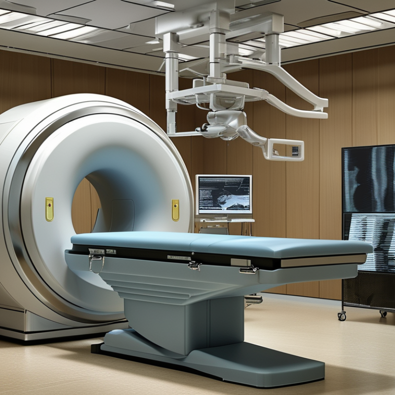
MRI artifacts become more frequent in bariatric patients because body size can challenge the scanner’s field, coils, and table design. The World Health Organization reported that more than one billion people lived with obesity in 2022. This growing need makes artifact control clinically important. Common problems include motion ghosting, signal loss near the body edge, truncation, and uneven fat suppression. Limited space may also force the patient’s arms against the torso, creating shading and coil-related intensity changes.
A radiographer should check the patient’s weight, girth, and comfort before scanning. The ACR Manual on MR Safety emphasizes confirming equipment limits and safe positioning before entry. Larger body diameter can reduce signal-to-noise ratio, especially when the anatomy extends beyond the receiver coil. Parallel imaging may shorten acquisition time, but aggressive acceleration can increase noise and residual artifacts. It is tempting to increase the field of view alone. That may reduce truncation, but it can also lower spatial detail. No protocol works perfectly.
Tips: Use the largest suitable coil and center the patient carefully. Add padding to reduce movement and pressure discomfort. Consider breathing instructions, faster sequences, and repeated localizer images. Inspect fat suppression across the entire field, not only the center. If shading remains, repositioning may help more than repeating the same sequence. Document the limitation clearly for the interpreting radiologist. A practical compromise is sometimes necessary. (Sources: WHO, World Obesity Report, 2024; ACR Manual on MR Safety, 2024.)
Bariatric MRI planning starts with equipment, not the scan sequence. In the United States, 40.3% of adults had obesity during 2021–2023, according to NCHS Data Brief No. 508 (2024). That figure makes capacity planning essential. Measure the patient’s widest point, then verify the table limit, bore diameter, and padding clearance. Do not rely on remembered specifications. Fit matters. A wide-bore system can improve access, but it may reduce gradient performance or available coils. The ACR Manual on MR Safety (2024) also emphasizes patient screening, safe positioning, and documented equipment limits.
Protocol design should protect signal-to-noise ratio while controlling scan time. Use dedicated flexible coils when available, place the anatomy near magnet isocenter, and avoid excessive distance from the receive elements. Higher bandwidth can reduce chemical-shift artifacts, although it may lower signal. Parallel imaging and compressed sensing can shorten breath-holds, but aggressive acceleration sometimes creates noisy or incomplete images. Adjust phase-encoding direction when body contour causes wraparound. Consider Dixon-based fat suppression if conventional fat suppression becomes uneven. Shorter echo trains may reduce blurring, yet they can extend acquisition time. The European Society of Radiology highlights patient-specific optimization as a core principle in its imaging quality guidance.
One protocol will not fit every body. That is where planning can fail. Technologists should test positioning before entering the bore, explain emergency communication clearly, and record the successful setup for future examinations. Recheck comfort after the localizer; small shifts can change coil performance and diagnostic quality.
Reducing MRI artifacts in bariatric patients often starts before sequence selection. In practice, positioning can matter more than another aggressive correction algorithm. Confirm table, bore, and coil limits before the patient enters. Explain the plan clearly. Anxiety can increase movement. Center the anatomy of interest at magnet isocenter, even when body contours make this difficult. Use broad, firm padding to reduce rocking and pressure points. Keep the spine neutral when possible. Avoid forcing the patient into a painful posture.
Coil placement deserves careful attention. Choose the largest suitable surface coverage, then place it close to the target anatomy. Do not leave a wide air gap. A small folded towel can support the coil, but it should not restrict breathing or create an uneven signal path. Secure cables without tight loops, and keep them outside the imaging region when appropriate. For abdominal studies, align the coil with the liver, pancreas, or pelvis rather than the table edge. Ask the patient to practice a comfortable breath-hold before scanning.
Breathe in.
Hold.
Relax.
For patients near equipment limits, a second technologist should verify centering and clearance. This simple check can prevent repeat imaging. I have found that more padding is not always better; excessive material can shift the coil and reduce stability. Recheck comfort after scout images, because patients may slide or rotate during setup. Review the scout for off-center anatomy, wraparound risk, and motion before longer sequences. If the image remains poor, document what changed. That record helps the next examination, although it may not solve every artifact.
Managing Motion, Field Inhomogeneity, and Signal Loss
Bariatric MRI requires more than simply increasing the field of view. Limited space can make positioning uncomfortable, which often causes shifting, breathing changes, or repeated motion. Explain the scan clearly before it starts. Use firm but comfortable supports around the abdomen and legs. Keep the patient warm, since shivering can create additional artifacts. Shorter sequences may improve cooperation, although they can reduce resolution. There is no perfect compromise.
Field inhomogeneity becomes more noticeable across a larger body habitus. Careful centering is essential, even when the anatomy cannot fit ideally inside the coil. Use appropriate shimming, higher receiver bandwidth, and artifact-reduction techniques when available. Increase signal averages only when necessary, because longer acquisitions may invite more movement. Signal loss may also result from poor coil coverage or excessive distance from the sensitive elements. Check anatomy coverage before repeating a full sequence.
Tips: Secure the patient gently. Practice breath-holding first. Use padding to reduce fatigue. Ask about pain before scanning. Review each image immediately. If an image looks poor, identify the cause before changing every parameter. A small adjustment in positioning may help more than extra averages. Local protocols should guide limits, equipment use, and safety screening. Technologists should document what worked and what failed, because imperfect scans can still improve future planning.
Bariatric MRI examinations demand careful image-quality decisions, not automatic rescanning. The World Health Organization reported that more than one billion people were living with obesity in 2022. This increases the need for practical, patient-centered MRI workflows. Larger body size can reduce signal-to-noise ratio, limit coverage, and create uneven fat suppression. Body contact with the bore may also produce shading or motion.
Check the margins.
Before repeating a sequence, review anatomy, coverage, motion, signal uniformity, ghosting, and geometric accuracy. These are core image-quality measures in the 2024 American College of Radiology MRI Accreditation Program. A sequence should be repeated when artifacts obscure a clinically important structure, not simply because the image looks imperfect. Repositioning the patient, widening the field of view, changing phase direction, or improving coil placement may help. Shorter breath-holds can also reduce respiratory motion.
Repeat selectively.
A practical approach is to inspect each sequence immediately after acquisition. If abdominal fat suppression fails near the lateral body wall, repeat that sequence rather than the entire examination. If motion affects only one phase, targeted repetition protects patient comfort and scanner time. The 2024 WHO obesity data also remind departments that demand may continue rising. Still, technical solutions are not universal. Parallel imaging can lower signal, and extra averages may lengthen scanning. We sometimes repeat too quickly, without identifying the artifact source. That deserves review. A documented decision, including why a sequence was accepted or repeated, supports consistent reporting and safer patient care.
| Artifact or Limitation | Typical Cause in Bariatric MRI | Image-Quality Check | Recommended Optimization | Repeat the Sequence When |
|---|---|---|---|---|
| Reduced signal-to-noise ratio (SNR) | Greater distance between the anatomy and the receive coil, limited coil coverage, and increased noise from a large field of view. | Confirm that the target organ, lesion, or vessel can be distinguished from background noise and that small clinically relevant structures remain visible. | Use the largest appropriate dedicated or flexible coil coverage, center the patient carefully, select a suitable field of view, and increase averages or acquisition time only when clinically justified. | Repeat if the diagnostic target is obscured by noise or if the radiologist cannot confidently assess the clinical question. |
| Wrap-around (aliasing) | The anatomy extends beyond the selected field of view, especially in the phase-encoding direction. | Look for anatomy appearing on the opposite side of the image or overlapping the region of interest. | Increase the phase field of view, use phase oversampling, change the phase-encoding direction when appropriate, or apply validated anti-aliasing methods. | Repeat if aliasing overlaps the organ, lesion, spine, vessels, or other structure required for interpretation. |
| Motion and respiratory artifact | Longer scan times, discomfort, difficulty maintaining position, involuntary movement, or respiratory motion. | Assess ghosting across the anatomy of interest, blurring of margins, and loss of alignment between repeated acquisitions. | Explain the sequence before scanning, improve padding and comfort, use respiratory triggering or breath-holding when feasible, shorten the acquisition, and use motion-robust or parallel-imaging techniques. | Repeat when ghosting or blurring changes lesion conspicuity, vascular assessment, organ margins, or measurement accuracy. |
| Susceptibility and geometric distortion | Air–tissue interfaces, bowel gas, metallic materials, and long echo-planar readouts can produce signal loss or displacement. | Compare the distorted region with anatomic landmarks and determine whether the artifact affects the diagnostic target. | Use a shorter echo time when appropriate, reduce echo-planar echo spacing, increase receiver bandwidth, and select a non-echo-planar or spin-echo-based sequence when clinically suitable. | Repeat with a less distortion-sensitive sequence if the target is displaced, partially absent, or falsely enlarged. |
| Coil-related shading or signal nonuniformity | Uneven sensitivity across a large body contour, incomplete coil coverage, or a coil positioned too far from the anatomy. | Check for gradual intensity changes that could mimic pathology or conceal a lesion. | Reposition the patient and coil, ensure the coil elements cover the entire region of interest, and use validated intensity-uniformity correction during reconstruction. | Repeat if nonuniformity prevents reliable comparison of tissue signal or obscures a focal abnormality. |
| Fat-suppression failure | Large field inhomogeneity, increased tissue-to-coil distance, and frequency-selective suppression methods that are sensitive to magnetic-field variation. | Look for residual bright fat, uneven suppression, or loss of signal in tissue that should remain diagnostically visible. | Use a more uniform fat-suppression method when available, consider short-tau inversion recovery for appropriate applications, and verify adequate magnetic-field homogeneity before acquisition. | Repeat if residual fat overlaps a lesion or if uneven suppression prevents interpretation of edema, inflammation, or enhancement. |
| Partial-volume artifact | Thicker slices, large voxels, and limited spatial resolution needed to maintain acceptable scan time or SNR. | Determine whether adjacent tissues are averaged together and whether lesion margins or small structures are indistinct. | Use thinner slices or smaller voxels when the clinical question requires higher spatial resolution; compensate with an appropriate acquisition time and SNR strategy. | Repeat a targeted high-resolution series when a lesion boundary, duct, vessel, or small anatomical structure cannot be characterized. |
| Poor contrast or low lesion conspicuity | Suboptimal sequence weighting, insufficient temporal resolution, incorrect timing, or reduced SNR. | Confirm that normal tissue contrast is appropriate for the body region and that the suspected abnormality is visible on at least one relevant plane or sequence. | Adjust repetition time, echo time, inversion time, flip angle, temporal resolution, or contrast timing according to the examination protocol and clinical indication. | Repeat only the affected sequence or phase when the clinical question remains unanswered and the limitation is correctable. |
| Incomplete or truncated anatomy | Field of view, table position, coil coverage, or patient positioning does not include the entire region of interest. | Verify coverage at the superior and inferior edges and confirm that the prescribed anatomy is present in every required plane. | Recenter the patient, expand the field of view, use multiple stations when appropriate, and prescribe slices from localizers that include the full target. | Repeat when any clinically relevant portion of the target anatomy is outside the field of view or not covered by the coil. |
| Specific absorption rate (SAR) or scan-time limitation | Higher body mass, increased tissue loading, repeated radiofrequency pulses, and longer or high-flip-angle sequences can increase heating and prolong the examination. | Review system safety messages, sequence availability, scan duration, and whether the examination meets the intended diagnostic objective. | Follow scanner operating limits, use lower flip angles or longer repetition times when appropriate, reduce redundant sequences, and prioritize sequences that answer the clinical question. | Repeat only if a safety-limited or interrupted acquisition is nondiagnostic and an approved alternative sequence can provide the required information. |
| Patient-table or bore fit concern | Body dimensions may exceed equipment limits or restrict safe positioning and coil placement. | Confirm manufacturer-stated table weight, bore clearance, coil fit, patient comfort, and the ability to maintain the required position safely. | Perform pre-scan screening, use appropriate padding and positioning aids, maintain communication, and select equipment and protocols within documented safety limits. | Do not repeat solely for image quality if safe positioning cannot be maintained; escalate for protocol review or alternative imaging when necessary. |
| Overall diagnostic quality | Several moderate artifacts may combine to reduce confidence even when no single artifact is severe. | Rate the study as diagnostic, limited, or nondiagnostic for the specific clinical question; assess coverage, motion, SNR, contrast, and anatomic conspicuity together. | Use a targeted repeat strategy rather than repeating the entire examination. Document the limiting factor and the sequence selected for correction. | Repeat when the expected diagnostic benefit outweighs additional scan time, patient burden, safety considerations, and the likelihood of obtaining improved images. |
: Good positioning can reduce motion and artifacts before scanning begins. Center the target anatomy near magnet isocenter. Avoid painful postures.
Broad, firm padding can reduce rocking, pressure points, and fatigue. Too much padding may shift the coil. More is not always better.
Use the largest suitable coil coverage. Keep it close to the target anatomy, without a wide air gap. Secure it gently.
Explain the scan before starting. Practice a comfortable breath-hold. Use short instructions: “Breathe in. Hold. Relax.”
Confirm table, bore, and coil limits before entry. A second technologist should verify centering and clearance. This simple check can prevent repeats.
Center the anatomy carefully and use appropriate shimming. Review coil coverage and distance from sensitive elements. Higher bandwidth may help.
Repeat it when artifacts hide clinically important anatomy. Check motion, coverage, signal uniformity, ghosting, and geometric accuracy first.
No. Repeat only the affected sequence when possible. A local adjustment may help more than extra averages. There is no perfect compromise.
Record positioning changes, coil placement, artifact causes, and accepted or repeated sequences. The record may not solve everything, but it supports better planning.
How to reduce artifacts in MRI scans for bariatric patients? Start by recognizing the most common challenges, including motion blur, limited field of view, magnetic field inhomogeneity, coil-related signal loss, and reduced image uniformity. Selecting MRI equipment with an adequate bore size, table weight capacity, and suitable coil coverage can improve comfort and signal reception. Imaging protocols should be adapted to the patient’s body habitus, using appropriate field of view, slice thickness, acceleration settings, and sequences that balance resolution with scan time.
Careful positioning is equally important. Center the patient accurately, provide stable support, and place coils as close as practical to the anatomy while maintaining comfort and safety. Technologists should minimize movement, optimize shimming, and monitor for areas of weak signal or distortion. After scanning, evaluate image quality systematically to confirm that the diagnostic region is fully covered and clearly visible. If artifacts compromise interpretation, repeat only the affected sequences with adjusted positioning, parameters, or motion-control strategies.